
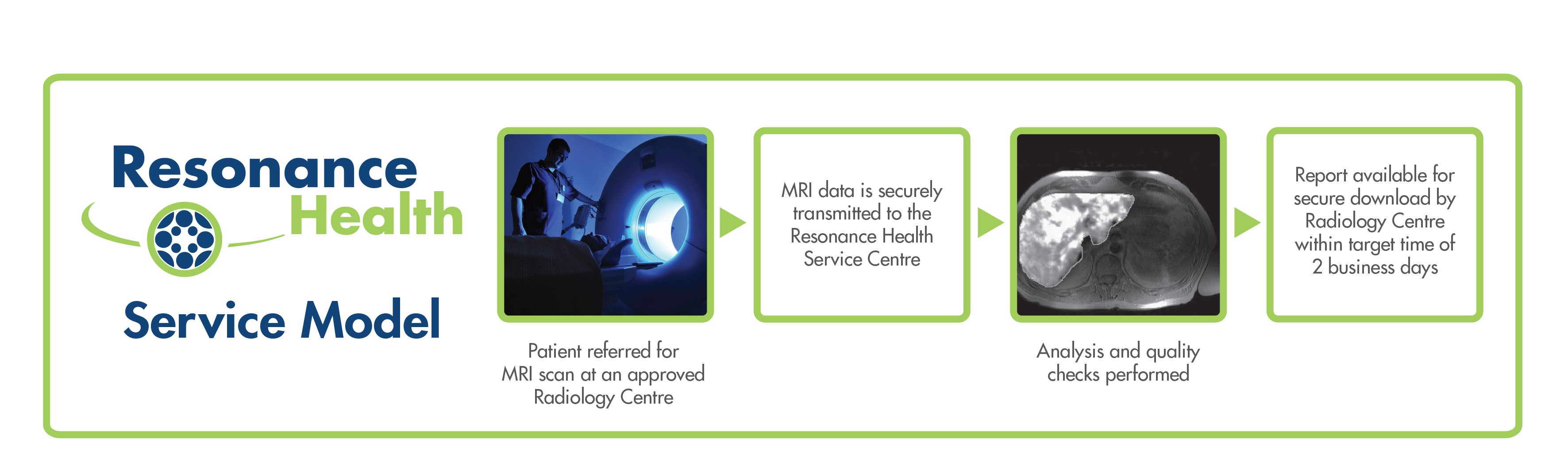
Following an easy setup process, Resonance Health provides a range of clinical image analysis services of MRI scans via a unique quality assured service delivery model:
- FerriScan® – the global gold standard for the measurement of liver iron concentration (LIC)
- Cardiac T2* – assessment of cardiac iron (FerriScan add-on)
- HepaFat-Scan® – measurement of volumetric liver fat fraction (VLFF)
- Bone Marrow R2-MRI – assessment of bone marrow iron
Image data is securely transferred to Resonance Health’s central ISO certified Service Centre, where it is analysed by our team of experts. Result reports are then made available for download by authorised Radiology staff.
- Services are charged per analysis only – there are no subscription or licence fees
- Radiology Centres are not required to purchase any additional hardware or software
- The imaging protocols require no contrast agents and can be established on most 1.5 Tesla scanners from Siemens, GE, and Phillips.
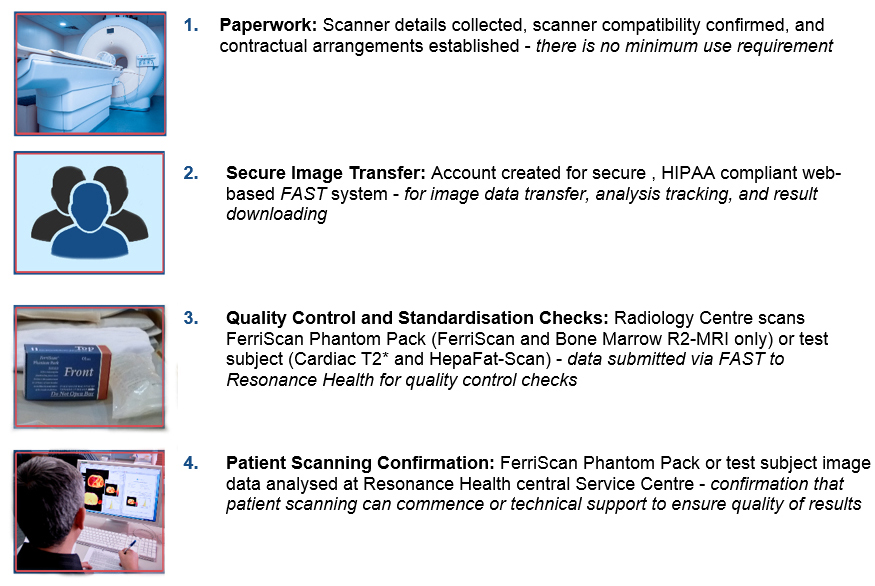
4 Step Easy Setup Process
To learn more about our services and how they can benefit your Radiology Centre, download our information sheets below.
- FerriScan Fact Sheet
- Easy Setup for Radiology
- FerriScan and Cardiac T2*Fact Sheet
- HepaFat-Scan Fact Sheet
- Bone Marrow R2-MRI Fact Sheet
Technical Requirements for Acquiring FerriScan and Bone Marrow Images
FerriScan requires an approximately 10 minute scan using a single spin echo sequence.
Minimum Radiology Centre requirements for the acquisition of suitable data for FerriScan and Bone Marrow R2-MRI:
- MRI scanner with a field strength of 1.5 Tesla
- Single spin echo sequence with a minimum TE of 6.0 ms
- A torso / chest / abdomen receiver coil
- The ability to transfer images to a networked computer with an internet connection
Technical Requirements for Acquiring Cardiac T2* Images
Cardiac T2* requires a single breath hold scan using a multi gradient echo sequence.
Minimum Radiology Centre requirements for the acquisition of suitable data for Cardiac T2*:
- Radiology Centre personnel with experience in acquiring cardiac images.
-
MRI scanner with a field strength of 1.5 Tesla and a cardiac MRI package that includes:
- Radio Frequency coil suitable for acquiring cardiac images
- ECG facility
-
Single breath-hold, multi-echo T2* sequence with:
- Total of 8 echo times
- Minimum TE between 2 and 3 ms
- Maximum TE between 16 and 23 ms
- The ability to transfer images to a networked computer with an internet connection
Technical Requirements for Acquiring HepaFat-Scan Images
HepaFat-Scan requires one to three single breath hold scans using a gradient echo sequence (the number of breath holds being dependent on the scanner model).
Minimum Radiology Centre requirements for the acquisition of suitable data for HepaFat-Scan:
- MRI scanner with a field strength of 1.5 Tesla
- Gradient echo sequence
- A torso / chest / abdomen receiver coil
- The ability to transfer images to a networked computer with an internet connection
If you are an established customer, click here to login to FAST